
Age-related respiratory decline is the progressive, measurable reduction in pulmonary function that begins in mid-adulthood, driven by structural changes in lung tissue, chest wall mechanics, and gas-exchange capacity. Research published in PMC documents the core benchmarks clinicians rely on: forced expiratory volume in one second (FEV1) falls at roughly 30 mL per year, while forced vital capacity (FVC) declines at approximately 20 mL per year in otherwise healthy adults. These losses are measured through spirometry, supplemented by diffusing capacity (DLCO) testing and pulse oximetry. The Merck Manual frames this as a predictable biological process, not a disease, though it creates the physiologic substrate on which respiratory diseases become more dangerous. Understanding these benchmarks is the first step toward knowing when normal aging ends and pathology begins.
At a glance: FEV1 declines approximately 30 mL/year; FVC approximately 20 mL/year; measurable decline typically begins around age 35.
Table of Contents
- How do lungs develop, and what baseline do clinicians measure against?
- What measurements show age-related respiratory decline?
- What structural and cellular changes drive the loss of function?
- How does respiratory aging affect daily function and exercise capacity?
- Which conditions become more common or more severe with age?
- What accelerates decline, and what can you do to slow it?
- When does decline begin, and how fast does it progress?
- When should you seek clinical evaluation?
- Medical oxygen vs. consumer portable oxygen: what you need to know
- Key Takeaways
- What the evidence actually tells us about respiratory aging
- Revo2 portable oxygen for short-term recovery and altitude support
- Useful sources and further reading
How do lungs develop, and what baseline do clinicians measure against?
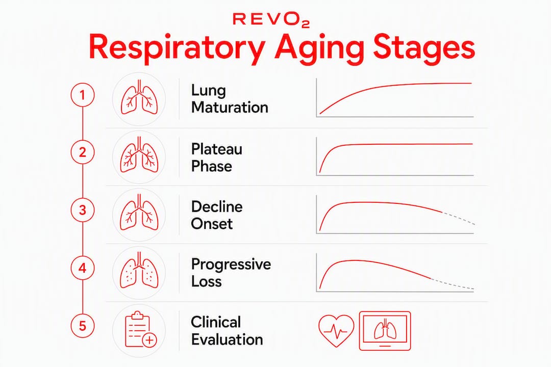
Lung development is not complete at birth. Alveolar multiplication continues through childhood, and the respiratory system reaches its structural and functional peak in early adulthood, typically between ages 20 and 25. Women tend to reach peak lung function slightly earlier than men, and absolute peak values are lower in women, partly because of smaller thoracic dimensions. After that peak, function plateaus briefly before the gradual decline begins.
This matters clinically because “normal” spirometry values are always expressed as a percentage of a predicted value derived from reference equations. Those equations account for age, sex, height, and, increasingly, ethnicity. Comparing a 70-year-old’s spirometry to a 25-year-old’s absolute values without adjustment would overestimate impairment. The reference equations used in the United States, such as those from the Global Lung Function Initiative (GLI), are built from large population datasets and produce age-specific predicted values.
Key maturation and baseline milestones:
- Peak FEV1 and FVC are typically reached between ages 20 and 25.
- Women reach peak function roughly 2–3 years earlier than men on average.
- Lung function plateaus briefly after peak, then begins a gradual decline around age 35.
- Height is the single strongest predictor of absolute lung volumes; taller individuals have larger predicted values.
- Ethnicity-specific reference equations exist because population-level differences in thoracic dimensions affect predicted values.
| Milestone | Typical Age | Clinical Implication |
|---|---|---|
| Peak lung maturation | 20–25 years | Establishes the individual’s reference baseline |
| Earlier peak in women | ~18–22 years | Women’s predicted values reflect smaller absolute volumes |
| Onset of measurable decline | ~35 years | Annual loss begins; longitudinal tracking becomes useful |
| Accelerated decline risk | 60+ years | Comorbidities and cumulative exposures amplify natural loss |
One limitation worth noting: most reference equations were built from predominantly white, non-Hispanic populations. Applying them to older adults from other ethnic backgrounds can produce systematically biased results, which is why clinicians increasingly use GLI multi-ethnic equations rather than older race-correction multipliers.
What measurements show age-related respiratory decline?
Spirometry is the primary tool. It measures how much air you can forcefully exhale and how fast, producing FEV1 (the volume exhaled in the first second), FVC (total exhaled volume), and the FEV1/FVC ratio. A ratio below 0.70 is the traditional threshold for airflow obstruction, though clinicians now prefer the lower limit of normal (LLN) approach, which accounts for the fact that the ratio naturally falls with age even in healthy lungs.
Beyond spirometry, DLCO measures how efficiently oxygen crosses from the alveoli into the bloodstream. Pulse oximetry gives a non-invasive estimate of arterial oxygen saturation. Cardiopulmonary exercise testing (CPET) quantifies maximal oxygen uptake (VO₂ max) and identifies whether exercise limitation is cardiac, pulmonary, or deconditioning-related.
| Measure | What It Captures | Age-Related Trend |
|---|---|---|
| FEV1 | Airflow in first second of forced exhalation | Declines ~30 mL/year after age 35 |
| FVC | Total forced exhaled volume | Declines ~20 mL/year after age 35 |
| FEV1/FVC ratio | Proportion of FVC exhaled in 1 second | Gradually falls; LLN preferred over fixed 0.70 cutoff |
| TLC | Total lung capacity | Relatively preserved; slight increase due to air trapping |
| RV | Residual volume (air remaining after full exhalation) | Increases with age as elastic recoil declines |
| DLCO | Diffusing capacity for carbon monoxide | Declines with reduced alveolar-capillary surface area |
| Resting PaO₂ | Arterial oxygen tension | Falls gradually; documented in PMC research |
Residual volume (RV) increases because aging lungs lose the elastic recoil needed to compress fully during exhalation. This means older adults trap more air at end-exhalation, which raises total lung capacity slightly even as usable ventilatory capacity falls.
Pro Tip: Clinicians use the LLN derived from age-adjusted reference equations rather than a fixed FEV1/FVC cutoff of 0.70. In a healthy 75-year-old, a ratio of 0.68 may be entirely normal. Using the fixed cutoff in older adults systematically overdiagnoses COPD.
What structural and cellular changes drive the loss of function?
The measurable numbers reflect real tissue-level changes. Elastic fibers in the lung parenchyma degrade over time, reducing the lung’s ability to recoil after inhalation. This decreased elastic recoil means the lung can inflate more easily (higher compliance) but cannot drive air out as forcefully, which directly explains the FEV1 decline. Small airways, no longer held open by the outward traction of surrounding elastic tissue, tend to collapse earlier during exhalation, trapping air and raising RV.
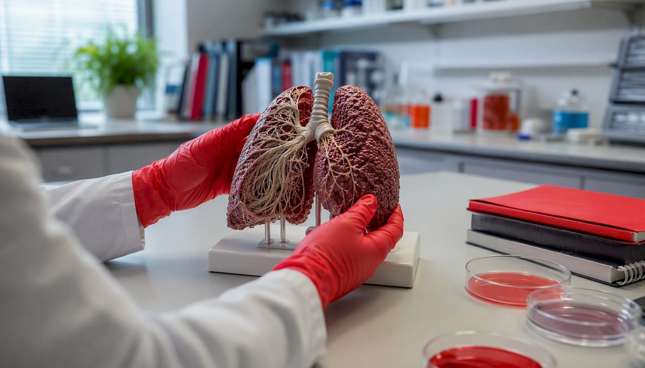
At the alveolar level, the picture is one of gradual simplification. Alveolar walls thin and merge, enlarging individual air spaces and reducing total surface area available for gas exchange. Frontiers research from 2024 documents that total alveolar surface area falls roughly 2.5 m² per decade between midlife and older ages, from approximately 70 m² in adults aged 30–39 to approximately 60 m² in adults aged 70–79. That loss of surface area, combined with thickening of the alveolar basement membrane and collagen deposition, directly reduces DLCO.
Structural changes in the aging lung:
- Loss of elastic fibers reduces recoil and allows small airway collapse.
- Alveolar enlargement and wall thinning reduce gas-exchange surface area.
- Increased anatomic dead space means a larger proportion of each breath ventilates airways rather than alveoli.
- Chest wall stiffening (calcification of costal cartilage, vertebral changes) increases the work of breathing.
- Diaphragm weakening and ribcage shape changes reduce ventilatory efficiency.
At the cellular level, the drivers are well-characterized. Oxidative stress accumulates as mitochondrial function declines, producing reactive oxygen species that damage structural proteins and DNA. Telomere shortening limits the replicative capacity of epithelial progenitor cells, impairing repair after injury. Chronic low-grade inflammation, sometimes called “inflammaging,” sustains a proinflammatory microenvironment that accelerates extracellular matrix remodeling.
“The aging lung faces a convergence of mechanical, cellular, and immunological stressors that individually are manageable but collectively erode the reserve capacity that protects against disease. Senescent cells accumulate, resist apoptosis, and secrete inflammatory mediators that further degrade the tissue environment — a process that is now a primary target of emerging anti-aging therapies.” — PMC review on lung aging biology
How does respiratory aging affect daily function and exercise capacity?
At rest, the changes described above are largely silent. A healthy 65-year-old sitting quietly will typically maintain normal oxygen saturation and will not feel short of breath. The functional ceiling, however, is meaningfully lower than it was at 35. This is the defining practical reality of respiratory aging: the reserve is compressed, not the baseline.
During moderate to vigorous exertion, the gap becomes apparent. Older adults reach their ventilatory limits at lower workloads, experience greater perceived breathlessness for the same absolute effort, and recover more slowly after stopping. VO₂ max declines with age partly because of cardiac output changes and partly because of the pulmonary limitations described above, including reduced DLCO and lower resting arterial oxygen tension.
Functional consequences of respiratory aging:
- Reduced maximal exercise capacity, with ventilatory limitation appearing at lower workloads.
- Greater perceived dyspnea on exertion relative to younger adults performing the same task.
- Slower recovery of breathing rate and perceived effort after physical activity.
- Reduced peak expiratory flow, which limits the speed of forced exhalation.
- Increased susceptibility to respiratory decompensation during acute illness, because the functional reserve is already narrowed.
Consider a practical scenario: an older adult who manages daily activities without difficulty may become significantly dyspneic when climbing two flights of stairs quickly or carrying heavy loads. After a respiratory infection, the temporary additional loss of function can push that person below the threshold for comfortable daily activity, even though their baseline was technically “normal for age.” Recovery from that infection takes longer because repair mechanisms are slower and the starting reserve was already reduced.
Which conditions become more common or more severe with age?
Normal respiratory aging does not cause disease by itself, but it lowers the threshold at which disease becomes serious. The Merck Manual and MedlinePlus both highlight how the combination of structural decline and immune senescence makes older adults disproportionately vulnerable to respiratory complications.
Age-associated respiratory conditions and risks:
- Pneumonia: Older adults face higher rates of pneumonia-related hospitalization and mortality. A diminished cough reflex, reduced mucociliary clearance, and impaired immune response all contribute. The reduced cough sensitivity is a particularly under-recognized risk factor because sputum and aspirated particles are less effectively cleared from the airways.
- COPD exacerbations: Age-related loss of elastic recoil overlaps mechanically with emphysema, making COPD more severe and harder to reverse in older patients.
- ARDS: Frontiers research links epithelial cell senescence and reduced repair capacity to worse outcomes and higher mortality in older patients with acute respiratory distress syndrome.
- Sleep-disordered breathing: Upper airway muscle tone declines with age, and chest wall stiffening alters breathing mechanics during sleep, increasing the prevalence of obstructive sleep apnea in older adults.
- Interstitial lung disease: Age-related fibrotic changes in the extracellular matrix create a permissive environment for progressive fibrosis.
Statistic: Older adults account for a disproportionate share of pneumonia-related deaths in the United States, with the risk rising steeply after age 65, according to CDC surveillance data.
Pro Tip: Older patients often present atypically. Pneumonia may appear as confusion or falls rather than classic fever and productive cough. Clinicians evaluating older adults for respiratory illness should compare findings to the patient’s own prior baseline rather than relying solely on age-group norms, because individual variability is wide.
What accelerates decline, and what can you do to slow it?
Not all respiratory aging is equal. Lifestyle and environmental exposures can dramatically accelerate the natural trajectory. Smoking is the largest modifiable accelerator, capable of multiplying the annual FEV1 loss several times over compared to a lifelong non-smoker. Long-term exposure to air pollution, occupational dusts, and recurrent respiratory infections each add cumulative damage that compounds the natural aging signal.
Primary accelerants of respiratory decline:
- Active and secondhand tobacco smoke (the dominant modifiable driver).
- Long-term exposure to particulate air pollution (PM2.5) and indoor combustion products.
- Occupational exposures: silica, asbestos, coal dust, and reactive chemicals.
- Recurrent lower respiratory infections, particularly in early life, which reduce peak lung function.
- Physical inactivity, which accelerates deconditioning and reduces respiratory muscle strength.
The good news is that several of these are modifiable, and the evidence for intervention is solid.
Evidence-based prevention checklist:
- Stop smoking. Smoking cessation at any age slows the accelerated FEV1 decline, though it does not fully restore lost function. Earlier cessation preserves more.
- Get vaccinated. Annual influenza vaccination, pneumococcal vaccination (PCV15 or PCV20 per current CDC recommendations), and RSV vaccination for adults 60 and older reduce the risk of infections that cause acute and lasting lung damage. Vaccination is specifically recommended because age-related oxidative stress and inflammation make older lungs more vulnerable to infectious insults.
- Maintain aerobic conditioning. Regular moderate-intensity aerobic exercise preserves VO₂ max, maintains respiratory muscle strength, and reduces perceived dyspnea. Pulmonary rehabilitation programs offer structured, supervised versions of this for those with established disease.
- Strengthen respiratory muscles. Inspiratory muscle training devices (threshold trainers) can improve respiratory muscle endurance, which is particularly relevant as diaphragm strength declines.
- Manage weight. Obesity increases the mechanical load on the respiratory system and worsens sleep-disordered breathing.
- Optimize nutrition. Adequate protein intake supports respiratory muscle mass. Antioxidant-rich diets (fruits, vegetables, omega-3 fatty acids) may reduce oxidative stress in lung tissue, though clinical trial evidence for specific supplements remains limited.
- Reduce indoor and outdoor pollution exposure. Use air filtration at home, avoid high-pollution outdoor activity days, and address indoor combustion sources such as gas stoves and wood-burning fireplaces.
When does decline begin, and how fast does it progress?
Lung maturation completes around age 20–25, followed by a brief plateau. Measurable spirometric decline typically begins around age 35 in healthy adults. The rates are gradual enough to be subclinical for decades, which is why many people first notice respiratory limitations only in their 50s or 60s, when cumulative loss finally crosses a functional threshold.
PMC research documents the core rates: FEV1 and FVC fall gradually each year in non-smoking healthy adults. DLCO also declines progressively, reflecting the alveolar surface area loss described above. Resting arterial oxygen tension (PaO₂) falls gradually, though resting oxygen saturation as measured by pulse oximetry typically remains above 95% in healthy older adults until advanced age.
| Measure | Approximate Rate of Change | Notes |
|---|---|---|
| FEV1 | ~30 mL/year | Accelerates significantly with smoking |
| FVC | ~20 mL/year | Decline rate varies by sex and height |
| Alveolar surface area | ~2.5 m²/decade | From ~70 m² (30s) to ~60 m² (70s) |
| Resting PaO₂ | Gradual decline | Remains clinically adequate in most healthy older adults |
| DLCO | Progressive decline | Reflects reduced alveolar-capillary surface |
Key figure: A non-smoking adult who reaches peak FEV1 of 4,000 mL at age 25 can expect, on average, a loss of roughly 1,500 mL by age 75 from aging alone, before any disease contribution.
Variability around these averages is substantial. Sex, height, ethnicity, comorbidities, and lifetime exposures all shift individual trajectories. Two 70-year-olds with identical demographics can have FEV1 values that differ by 30% or more. This heterogeneity is why clinicians track individual longitudinal trends in spirometry rather than relying solely on single cross-sectional comparisons to population norms.
When should you seek clinical evaluation?
Respiratory aging is normal, but certain symptoms signal that something beyond normal aging is happening. The distinction matters because treatable conditions, including COPD, interstitial lung disease, heart failure, and sleep apnea, can masquerade as “just getting older.”
Red-flag symptoms that warrant evaluation:
- New or progressive shortness of breath, particularly at rest or with minimal exertion.
- Resting oxygen saturation below 92% on room air, measured by pulse oximetry.
- Unexplained chronic cough lasting more than 8 weeks, or a change in sputum character or volume.
- Frequent respiratory infections (more than two lower respiratory infections per year).
- Waking unrefreshed, loud snoring, or witnessed apneas (suggesting sleep-disordered breathing).
- Unexplained weight loss combined with respiratory symptoms.
Diagnostic pathway for older adults:
Spirometry is the first-line test for airflow limitation. DLCO testing identifies diffusion impairment when spirometry is normal but symptoms persist. Chest imaging (X-ray or CT) evaluates parenchymal disease, masses, and pleural abnormalities. Overnight pulse oximetry or a formal polysomnography sleep study addresses suspected sleep apnea. CPET is reserved for cases where the cause of exercise limitation remains unclear after initial testing.
Clinicians interpreting results in older adults should use age-adjusted reference equations and compare findings to the patient’s own prior spirometry when available. A single test result is far less informative than a trend over time.
Medical oxygen vs. consumer portable oxygen: what you need to know
This distinction matters, and conflating the two can lead to real harm.
Prescription oxygen is indicated for chronic hypoxemia, defined as resting PaO₂ below 55 mmHg or oxygen saturation at or below 88% on room air, confirmed on two measurements. It is a medical treatment, prescribed by a clinician, titrated to a target saturation, and delivered through regulated equipment. It is not interchangeable with recreational products.
Consumer portable canned oxygen, such as the products Revo2 offers, is a different category entirely. These products deliver short-term supplemental oxygen for uses like altitude support, post-exercise recovery, brief fatigue relief, and general wellness. They are not intended to treat hypoxemia or replace prescribed oxygen therapy. Understanding the difference between oxygen therapy and supplemental oxygen is important before making any purchasing decision.
Appropriate uses for consumer portable oxygen:
- Short-term support at altitude, where ambient oxygen partial pressure is reduced.
- Brief post-exercise recovery support for athletes or active seniors.
- Situational fatigue or stress relief in otherwise healthy individuals.
- Supplemental use during cooldown after vigorous physical activity.
Safety considerations:
- Never use canned oxygen near open flames or in enclosed spaces with ignition sources; oxygen accelerates combustion.
- Consumer portable oxygen is not a substitute for prescribed medical oxygen in individuals with documented hypoxemia.
- If you experience resting oxygen saturation below 92%, consult a clinician before relying on any consumer product.
Pro Tip: Revo2’s safe-use guide walks through proper inhalation technique with the zero-leak mouthpiece, which minimizes waste and maximizes the benefit of each breath. Reviewing it before first use is worthwhile.
For older adults interested in how supplemental oxygen may support active living, Revo2’s guide to oxygen and senior mobility offers a science-backed overview of the evidence and appropriate use cases.
Key Takeaways
Age-related respiratory decline is a measurable, predictable process beginning around age 35, driven by structural and cellular changes that reduce FEV1, FVC, and gas-exchange capacity, and it is substantially modifiable through lifestyle choices and clinical monitoring.
| Point | Details |
|---|---|
| Decline begins around age 35 | FEV1 and FVC decline steadily in healthy non-smokers after this age. |
| Structural changes drive the numbers | Loss of elastic recoil, alveolar surface area reduction (~2.5 m²/decade), and chest wall stiffening all contribute. |
| Smoking is the top modifiable risk | Tobacco smoke accelerates FEV1 loss several times beyond the natural rate; cessation at any age slows further decline. |
| Vaccination and exercise are protective | Annual flu shot, pneumococcal vaccine, and regular aerobic conditioning are the highest-yield prevention steps. |
| Revo2 for short-term support | Revo2’s 98% pure canned oxygen is designed for altitude, post-exercise recovery, and brief fatigue relief, not as a substitute for prescribed medical oxygen. |
What the evidence actually tells us about respiratory aging
The conventional framing of respiratory aging as a slow, inevitable slide tends to obscure two things that the evidence makes clear. First, the rate of decline is far more variable than the average numbers suggest. A 70-year-old who has never smoked, exercises regularly, and has avoided significant pollution exposure may have lung function that rivals someone 15 years younger. The average decline rates are population statistics, not individual destinies. Second, the clinical community has historically underestimated how much of what gets labeled “normal aging” is actually accumulated exposure damage. When you strip out smoking, pollution, and recurrent infections from population datasets, the residual aging signal is real but modest.
What this means practically: the most important conversation an older adult can have about respiratory health is not “how much have I lost?” but “what am I doing that is accelerating loss, and what can I do to slow it?” The structural changes are not reversible, but the trajectory is. Aerobic conditioning preserves the functional reserve that determines whether a respiratory infection becomes a hospitalization or a week of feeling unwell. Vaccination reduces the acute insults that cause lasting damage. These are not minor interventions. They are the difference between aging with adequate respiratory reserve and aging without it.
Revo2 portable oxygen for short-term recovery and altitude support
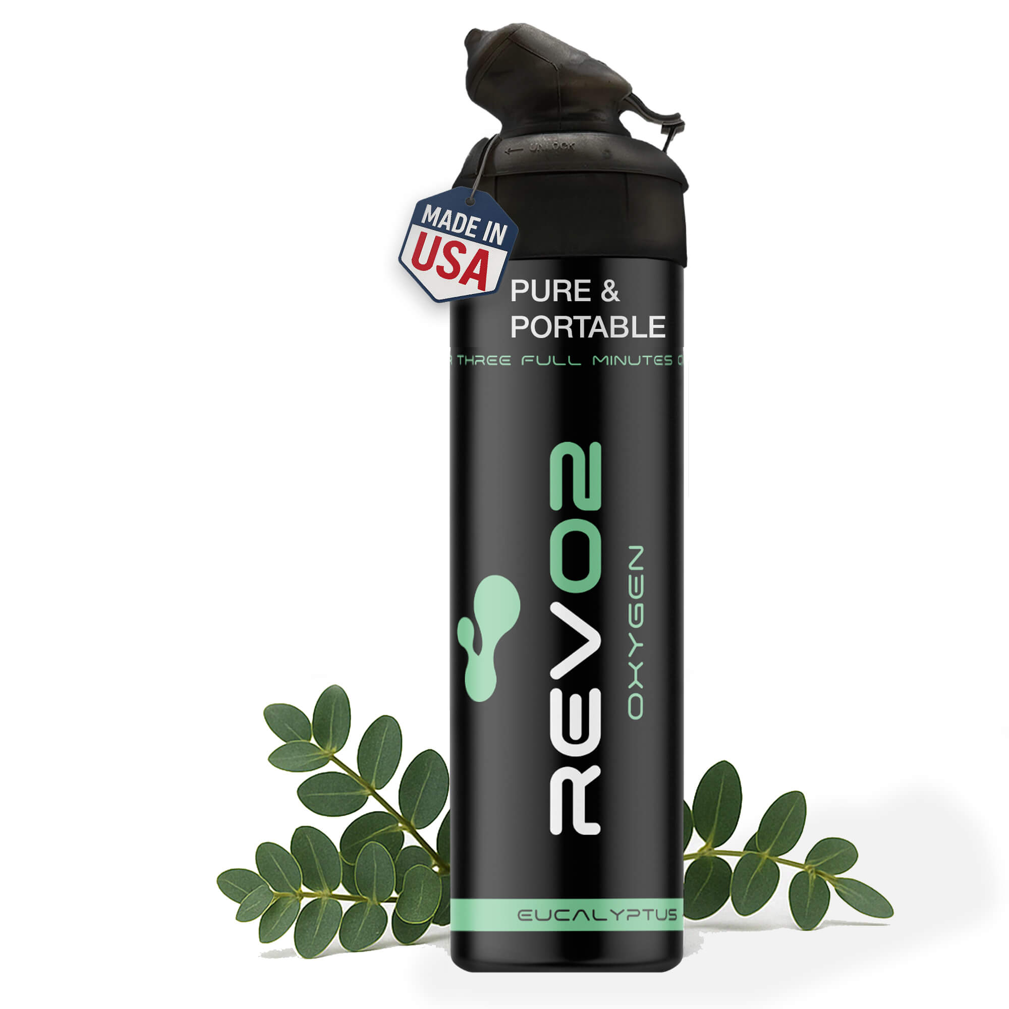
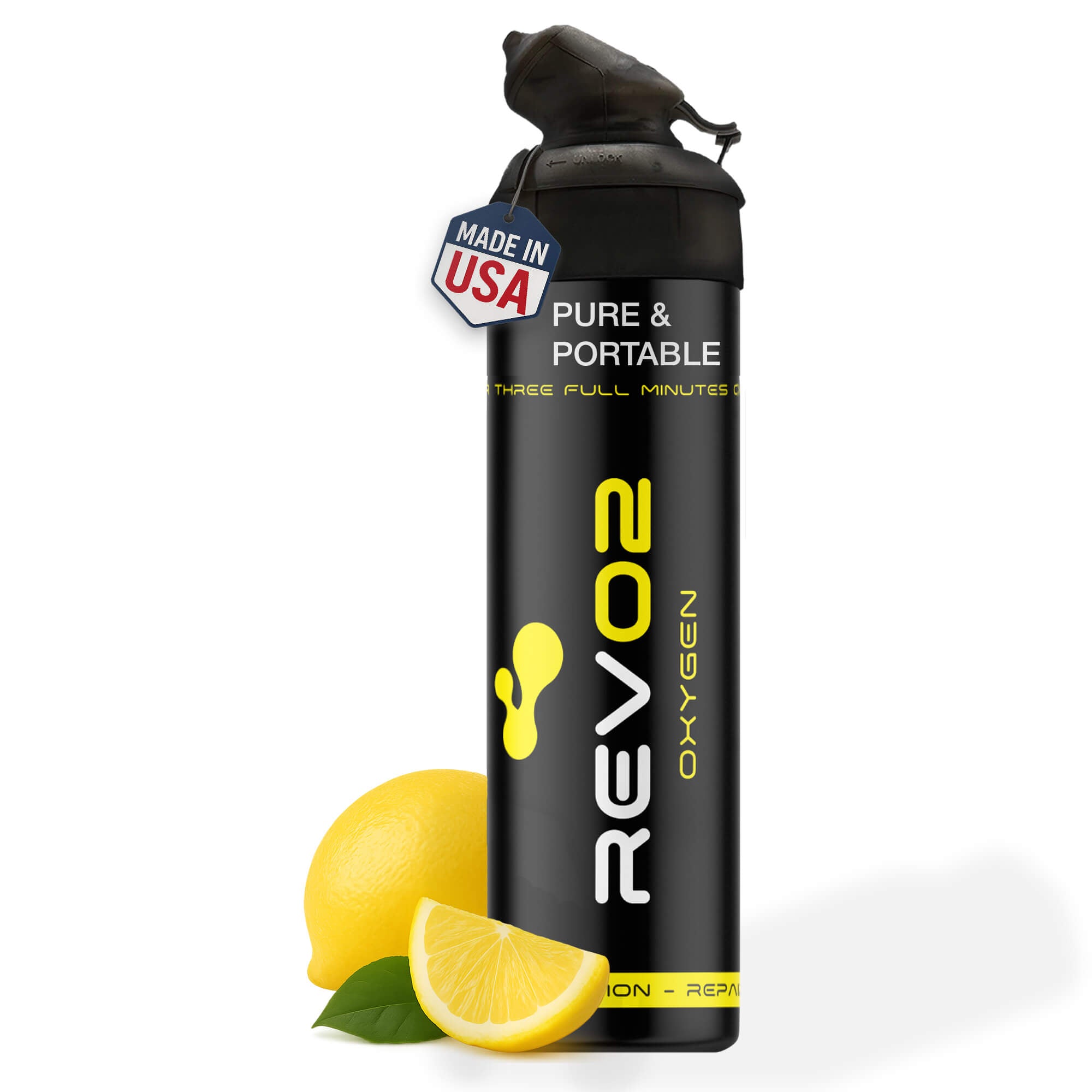
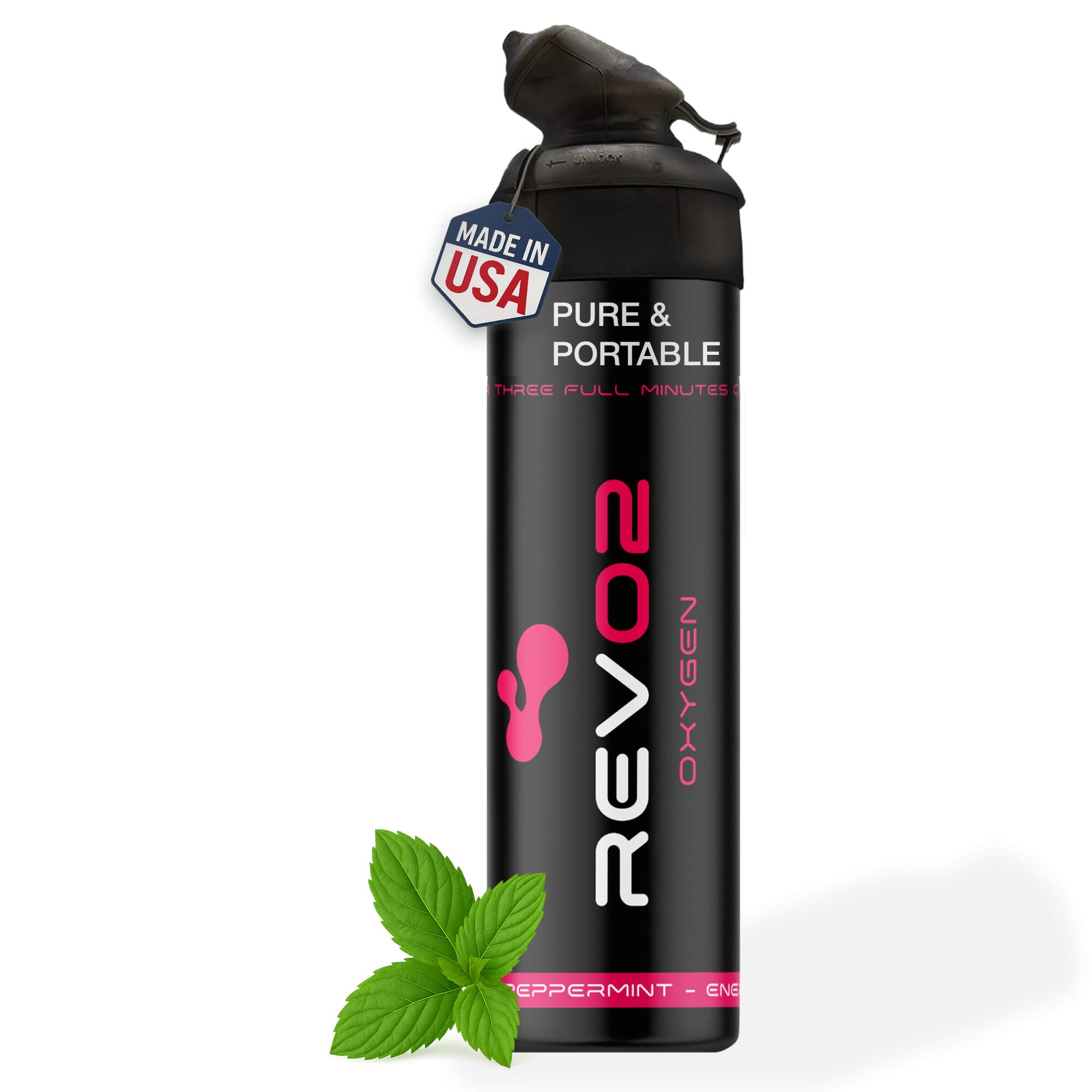
For older adults, athletes, and anyone navigating the real-world effects of reduced respiratory reserve, Revo2 offers a practical, portable option for short-term oxygen support. Unlike prescription oxygen systems that require clinical setup and ongoing medical management, Revo2’s canned oxygen collection delivers 98% pure oxygen through a zero-leak mouthpiece, with no mask, no prescription, and no complicated equipment. It is designed for the moments when you want a quick recovery boost after exertion, need support at altitude, or simply want to clear the mental fog that comes with fatigue.

Revo2 is not a medical treatment and is not intended for individuals with diagnosed hypoxemia or chronic respiratory disease requiring prescription oxygen. It is a wellness and performance adjunct for healthy adults who want accessible respiratory support in everyday situations.
Intended uses:
- Post-exercise recovery and cooldown support.
- Altitude adjustment for hikers, travelers, and skiers.
- Brief fatigue or stress relief during demanding days.
Revo2’s peppermint, lemon, and eucalyptus blends are available individually or in multi-packs. Browse the full canned oxygen collection to find the format that fits your routine.
This article provides general educational information about respiratory physiology and aging. It is not a substitute for professional medical advice, diagnosis, or treatment. Consult a qualified clinician for personalized guidance, especially if you are experiencing respiratory symptoms or abnormal test results.
Useful sources and further reading
- Aging and Lung Disease (PMC, PMC7998901) — Primary source for FEV1/FVC decline rates, gas-exchange changes, and longitudinal tracking guidance.
- The aging lung: Physiology, disease, and immunity (PMC, PMC8052295) — Comprehensive review of cellular and immunological mechanisms underlying lung aging.
- Aging changes in the lungs (MedlinePlus) — NLM-reviewed overview of chest wall, diaphragm, cough reflex, and immune changes in older adults.