
Physiologically, burnout is a state in which the body’s stress-response systems fail to return to baseline after chronic work stress, producing measurable autonomic and neuroendocrine changes. The core markers researchers and clinicians track include salivary cortisol diurnal patterns, vagally-mediated heart rate variability (HRV), particularly the RMSSD metric, and inflammatory markers such as C-reactive protein (CRP) and interleukin-6 (IL-6). Understanding burnout physiology explained through these measurable signals matters because it shifts the conversation from “you just need a vacation” to “your nervous system has been structurally altered by sustained overload.”
Key physiological signals associated with burnout include:
- Cortisol dysregulation: Flattened or blunted diurnal cortisol patterns, sometimes shifting from early hyperreactivity to later hyporeactivity as burnout deepens
- Reduced vagal tone: Lower RMSSD and high-frequency HRV power, indicating the parasympathetic nervous system is losing its restorative capacity
- Systemic inflammation: Elevated CRP and IL-6 levels, linking chronic stress to immune and cardiovascular risk
- Autonomic imbalance: Sympathetic nervous system dominance early, followed by overall autonomic dysregulation in chronic stages
One honest caveat upfront: findings across studies are heterogeneous, and burnout shares enough biomarkers with major depressive disorder that distinguishing the two purely by physiology remains an active research challenge. The biology of burnout involves real, measurable changes, but no single biomarker is diagnostic on its own.
Key Takeaways
Burnout is a measurable physiological syndrome driven by HPA-axis dysregulation and chronic suppression of vagal tone, with the emotional exhaustion component showing the most consistent biological associations.
| Point | Details |
|---|---|
| HPA axis dysregulation | Burnout progresses from early cortisol hyperreactivity to later hyporeactivity as chronic stress depletes the stress-response system. |
| Vagal tone suppression | Reduced RMSSD and HF-power HRV are the most consistently measured autonomic markers, linked to both exhaustion severity and cardiovascular risk. |
| Neurobiological changes | Prefrontal cortex, hippocampus, and amygdala all show functional and structural changes that explain cognitive fog, memory issues, and emotional dysregulation. |
| Burnout vs. depression overlap | Both conditions share reduced HRV and HPA alterations; emotional exhaustion drives most physiological associations, and comorbid depression complicates recovery. |
| Revo2 portable oxygen | Revo2’s 98% pure canned oxygen offers short-term breathing support for acute fatigue or stress moments; it is not a burnout treatment but a practical adjunct tool. |
Table of Contents
- What is burnout physiology explained through the Maslach framework?
- How does chronic work stress alter your HPA axis and autonomic nervous system?
- What brain changes explain the cognitive and emotional symptoms of burnout?
- Which biomarkers do researchers actually measure in burnout studies?
- What physical symptoms does burnout cause, and what are the long-term health risks?
- How does burnout differ from depression, and where do they overlap?
- Why does recent research focus so heavily on vagal function and HRV?
- Which interventions actually restore physiological regulation?
- When should you see a clinician, and what should you tell them?
- The physiology-first framing changes everything about how we treat burnout
- Revo2 portable oxygen as a short-term breathing support tool
- Sources
What is burnout physiology explained through the Maslach framework?
Burnout is not simply exhaustion from a hard week. Clinically, it is defined through the Maslach framework as a three-dimensional occupational syndrome: emotional exhaustion (the depletion of emotional resources), depersonalization or cynicism (a detached, indifferent, or negative stance toward work and colleagues), and reduced professional efficacy (a declining sense of competence and accomplishment). The Maslach Burnout Inventory (MBI) remains the most widely used standardized tool for operationalizing these dimensions in both research and clinical settings.
The progression matters for physiology because each stage carries a different biological signature. Early burnout typically looks like hyperarousal: the sympathetic nervous system is running hot, cortisol output is elevated, and the body is mobilizing resources it cannot fully replenish. This is the “fight or flight” system stuck in the on position. Over months or years of sustained occupational demand without adequate recovery, the system shifts. The HPA axis, which governs the cortisol stress response, begins to show signs of wear. Output can drop below normal, a pattern called hyporeactivity, and the parasympathetic nervous system loses its ability to restore calm.
The concept of allostatic load captures this wear-and-tear process well. Allostasis refers to the body’s ability to achieve stability through change, adjusting heart rate, cortisol, blood pressure, and immune function in response to demands. When those demands are chronic and recovery is insufficient, allostatic load accumulates across multiple systems simultaneously. The physiological costs are not confined to one organ or hormone; they spread across the cardiovascular, endocrine, immune, and nervous systems together.
Workplace demand-resource imbalance sits at the root of this cascade. When job demands consistently outpace the resources available (autonomy, social support, adequate rest, fair compensation), the body treats the gap as a chronic threat. That sustained threat perception is what drives the physiological progression from early hyperarousal to later exhaustion and hyporeactivity.
How does chronic work stress alter your HPA axis and autonomic nervous system?
The stress-response cascade begins in the hypothalamus. Perceived threat triggers the release of corticotropin-releasing hormone (CRH), which signals the pituitary gland to release adrenocorticotropic hormone (ACTH), which in turn drives the adrenal cortex to produce cortisol. Simultaneously, the sympathetic nervous system releases catecholamines, primarily adrenaline (epinephrine) and noradrenaline (norepinephrine), preparing the body for immediate action. This is the acute stress response, and it is adaptive in short bursts.
Chronic occupational stress keeps this system activated far beyond its design parameters. The staged cascade model, supported by review evidence on HPA and ANS function in burnout, describes the progression this way:
- Early phase: Elevated sympathetic tone and HPA hyperactivity; cortisol output is high, heart rate is elevated, and the body is in a state of sustained mobilization
- Intermediate phase: Regulatory feedback begins to break down; cortisol patterns flatten, and the normal morning cortisol awakening response becomes blunted
- Chronic/severe phase: HPA hyporeactivity sets in; cortisol output drops below normal; parasympathetic (vagal) tone is reduced, leaving the body unable to shift into restorative mode even during rest
The parasympathetic nervous system, mediated largely through the vagus nerve, is responsible for the “rest and digest” state that allows the body to repair, consolidate memory, regulate digestion, and restore cardiovascular function. When vagal tone is chronically suppressed, none of those restorative processes work efficiently. The 2021 review on the biology of burnout concluded that burnout is associated with sustained autonomic activation, HPA-axis alterations, and increased allostatic load, while also emphasizing that findings are heterogeneous and longitudinal evidence remains limited.
Allostatic load as a unifying framework is useful here because it explains why burnout does not feel like one thing. The body is paying costs across multiple systems at once: elevated inflammatory markers, disrupted sleep architecture, altered glucose regulation, and reduced immune surveillance. Each system compounds the others in a vicious cycle where poor sleep raises cortisol, elevated cortisol disrupts sleep, and reduced vagal tone makes both worse.
What brain changes explain the cognitive and emotional symptoms of burnout?
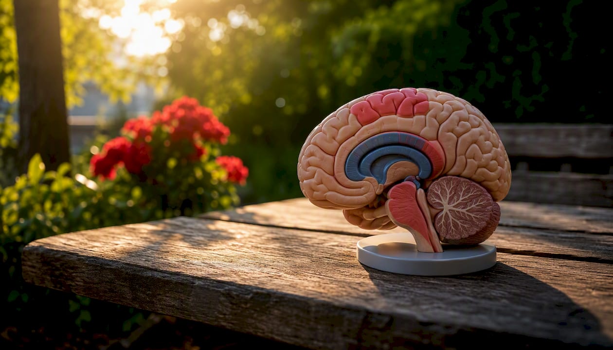
The neurobiological picture of burnout centers on three regions: the prefrontal cortex (PFC), the hippocampus, and the amygdala. Each plays a distinct role in stress regulation, and each shows evidence of functional or structural change under chronic stress conditions. Neuroscience summaries confirm that prolonged burnout correlates with impaired cognitive performance and measurable changes in brain function, though many studies are cross-sectional and cannot establish causation.
Prefrontal cortex: executive function under siege
The PFC governs working memory, decision-making, attention regulation, and emotional control. Chronic stress reduces PFC activity and connectivity, which maps directly onto the cognitive symptoms people with burnout describe: difficulty concentrating, poor decision-making, an inability to plan or prioritize, and a sense that mental tasks that once felt easy now require enormous effort. Neuroimaging studies have found reduced gray matter volume and altered activation patterns in PFC regions in individuals with high burnout scores, though sample sizes in most studies are small.
Hippocampus: memory and stress regulation
The hippocampus is particularly sensitive to cortisol. Prolonged cortisol elevation is associated with reduced hippocampal volume and impaired neurogenesis, the process by which new neurons form. This matters because the hippocampus plays a key role in both memory consolidation and in providing negative feedback to the HPA axis, helping shut down the cortisol response after a stressor passes. When hippocampal function is compromised, the HPA axis loses some of its braking capacity, which can perpetuate the cortisol dysregulation cycle.
Amygdala: threat detection in overdrive
The amygdala processes emotional salience and threat. Under chronic stress, amygdala reactivity tends to increase while PFC-amygdala connectivity weakens. The practical result is that emotional responses become harder to regulate: minor frustrations feel disproportionately threatening, emotional detachment or cynicism may develop as a defensive response, and the capacity for positive emotional engagement at work diminishes. This maps onto the depersonalization dimension of the Maslach framework.
A critical limitation worth naming: most neuroimaging studies in burnout use cross-sectional designs with relatively small samples, making it impossible to determine whether brain changes precede burnout, result from it, or reflect a shared underlying vulnerability. Longitudinal neuroimaging studies with adequate controls are still needed before causal claims can be made with confidence.
Which biomarkers do researchers actually measure in burnout studies?
Objective measurement is what separates burnout research from self-report alone. Several biological markers have been studied as potential indicators of physiological burnout, each with distinct strengths and limitations.
| Biomarker | What it measures | Typical finding in burnout | Key limitations |
|---|---|---|---|
| Diurnal salivary cortisol | HPA-axis activity across the day | Blunted morning awakening response; flattened diurnal slope in chronic burnout | Highly sensitive to sampling time, sleep quality, caffeine, and smoking |
| RMSSD (HRV metric) | Vagally-mediated parasympathetic tone | Reduced at rest and during stress tasks in high-exhaustion groups | Affected by age, BMI, fitness level, and measurement protocol |
| HF-power (HRV metric) | High-frequency vagal modulation | Reduced in burnout samples, especially in exhaustion-dominant cases | Requires controlled recording conditions; body position matters |
| CRP (C-reactive protein) | Systemic inflammation | Modestly elevated in some burnout samples | Non-specific; elevated by many conditions including infection and obesity |
| IL-6 (interleukin-6) | Pro-inflammatory cytokine signaling | Elevated in some chronic stress and burnout studies | Diurnal variation; confounded by physical activity and BMI |
The PMC population-based study found that emotional exhaustion was inversely associated with RMSSD after controlling for age, sex, BMI, substance use, and exercise, making it one of the more methodologically careful demonstrations of vagal suppression in burnout. Still, the authors caution against causal interpretation and note that depressive somatic symptoms attenuated some of the associations.
Recent empirical work published in Frontiers in Psychiatry has expanded the biomarker picture, examining neuroendocrine and autonomic measures across multiple populations, but results remain mixed. The Frontiers in Human Neuroscience review on autonomic markers similarly highlights vagal dysfunction as a promising mechanistic link to cardiovascular risk while acknowledging varied outcomes across studies.
Pro Tip: If you are tracking your own stress physiology, consumer-grade wearables that report RMSSD (such as Garmin, Polar, or WHOOP devices) can give you a directional sense of your vagal tone over time. They are not clinical instruments, but a sustained downward trend in RMSSD alongside work stress is worth discussing with a clinician.
What physical symptoms does burnout cause, and what are the long-term health risks?
The physiological aspects of burnout are not confined to lab values. They show up in the body in ways that often bring people to a doctor before anyone has named burnout as the underlying cause.
Common somatic manifestations include persistent fatigue that sleep does not resolve, tension headaches, gastrointestinal complaints such as heartburn and irritable bowel symptoms, muscle tension particularly in the neck and shoulders, and disrupted sleep architecture including difficulty falling asleep or staying asleep despite feeling exhausted. One clinical study reported headaches in 89% of burnout patients, suggesting the symptom often precedes formal occupational recognition of the syndrome. That figure underscores how frequently burnout presents as a physical complaint rather than a psychological one.
The long-term health associations are more sobering. Chronic HPA-axis dysregulation and sustained sympathetic activation are linked to elevated cardiovascular risk through several pathways: higher resting blood pressure, pro-inflammatory cytokine activity, altered lipid metabolism, and reduced heart rate variability. The 2021 biology of burnout review identifies immune changes as another downstream consequence, and clinical reports describe higher infection frequency and longer recovery times in chronic burnout samples, consistent with HPA-driven immune suppression.
The relationship between chronic stress and low oxygen levels is another physiological thread worth noting. Sustained sympathetic activation alters breathing patterns, often producing shallow, rapid respiration that reduces effective oxygen exchange. Over time, this can contribute to the fatigue and cognitive fog that burnout patients describe.
What makes the somatic picture clinically tricky is that each symptom can be attributed to a different cause: the headaches to tension, the GI symptoms to diet, the fatigue to poor sleep. Burnout as the unifying explanation often goes unrecognized until the pattern is viewed as a whole.
How does burnout differ from depression, and where do they overlap?
The physiological overlap between burnout and major depressive disorder (MDD) is substantial enough that researchers have debated whether burnout is a distinct clinical entity or a work-specific variant of depression. Understanding where they converge and where they diverge matters for both treatment and for interpreting biomarker findings.
Areas of overlap:
- Both conditions feature reduced vagally-mediated HRV and autonomic dysregulation
- Both show altered HPA-axis markers, including blunted cortisol awakening responses
- Both involve persistent fatigue, sleep disruption, and cognitive impairment
- Inflammatory markers (CRP, IL-6) are elevated in both conditions
Distinguishing features:
- Burnout is definitionally tied to occupational context; symptoms typically improve with sustained removal from the work environment, at least initially
- The core of burnout is emotional exhaustion and cynicism/depersonalization directed at work, not the pervasive low mood and anhedonia (loss of pleasure in all activities) that characterize MDD
- Depression involves a broader loss of motivation and pleasure that extends across all life domains, not just work
- Suicidal ideation and severe psychomotor changes are more characteristic of MDD than of burnout alone
The PMC population-based study provides a nuanced finding here: depressive somatic symptoms attenuated some of the associations between emotional exhaustion and reduced RMSSD, suggesting that when burnout and depression co-occur, the physiological signal becomes harder to parse. The exhaustion subcomponent of burnout appears to drive many of the autonomic and HPA associations, while depersonalization and reduced efficacy show weaker or less consistent physiological correlates.
Clinically, the two conditions frequently co-occur, and comorbid depression in a burnout patient tends to worsen physiological dysregulation and complicate recovery. Treating the occupational stressors without addressing the depressive component, or vice versa, typically produces incomplete results.
Why does recent research focus so heavily on vagal function and HRV?
Heart rate variability has become one of the most active measurement targets in burnout research for a straightforward physiological reason: RMSSD and high-frequency (HF) power are noninvasive, quantifiable indices of vagal tone, and vagal tone is the primary mechanism through which the parasympathetic nervous system restores cardiovascular and metabolic homeostasis after stress. A body that cannot upregulate vagal activity after a stressor cannot recover efficiently, and that failure of recovery is central to what burnout physiology actually is.
The PMC study remains one of the stronger demonstrations of this link, showing that emotional exhaustion was associated with lower RMSSD in a large, heterogeneous population sample after controlling for major confounders. The Frontiers in Human Neuroscience review on autonomic markers in stress-related conditions found mixed results across HRV studies but consistently identified vagal dysfunction as a correlate of exhaustion and a plausible mechanistic link to cardiovascular risk. Work published in Frontiers in Psychiatry similarly expanded the biomarker picture while acknowledging that inconsistent findings across populations make definitive conclusions premature.
Several research gaps remain clear. Most HRV studies in burnout are cross-sectional, meaning they capture a snapshot rather than tracking how vagal tone changes as burnout develops and resolves. Measurement protocols vary widely: recording duration, body position, time of day, and whether participants are at rest or under a stress task all affect RMSSD values, making cross-study comparisons difficult. Sample sizes in neuroimaging and HRV studies are often small. What the field needs most are well-controlled longitudinal studies that follow participants from early stress exposure through burnout onset and into recovery, measuring HRV and HPA markers at multiple time points.
Which interventions actually restore physiological regulation?
Effective burnout recovery targets the physiological systems that have been disrupted: vagal tone, HPA-axis regulation, inflammatory load, and sleep architecture. The evidence base varies considerably by intervention type.
Interventions with the strongest physiological rationale and evidence:
- Cognitive Behavioral Therapy (CBT): Addresses the cognitive appraisal patterns that sustain threat perception and HPA activation. Multiple trials show reductions in burnout scores and improvements in sleep; some studies report modest HRV improvements. CBT is the most evidence-supported psychological intervention for burnout.
- Mindfulness-Based Stress Reduction (MBSR): Directly targets autonomic regulation through breath-focused attention and body awareness. Studies show increases in HRV and reductions in cortisol in stressed populations, though burnout-specific longitudinal data are limited.
- Aerobic exercise: One of the most reliable vagal tone enhancers available. Regular moderate-intensity aerobic activity (150 minutes per week is the commonly cited threshold) consistently increases RMSSD and reduces inflammatory markers including CRP and IL-6. Effects on burnout-specific symptoms are supported by multiple trials.
- Sleep optimization: Sleep is the primary window for HPA-axis recovery and hippocampal consolidation. Interventions targeting sleep hygiene, including consistent sleep timing, reduced screen exposure before bed, and cognitive techniques for pre-sleep arousal, address one of the most physiologically damaging aspects of burnout.
- Workplace and organizational change: Individual-level interventions have limited durability when the occupational stressors driving allostatic load remain unchanged. Experts emphasize that systemic workplace improvements, including workload management, increased autonomy, and social support structures, are necessary to prevent physiological cascades from recurring. Without organizational change, individual recovery strategies are often temporary.
Realistic expectations matter here. Physiological markers like HRV and cortisol patterns can begin to shift within weeks of consistent intervention, but full normalization of HPA function in severe, chronic burnout may take months. The Psychology Today overview notes that practitioners commonly recommend interventions that restore autonomic balance and agency, including behavioral activation and activities that rebuild a sense of control, alongside formal psychological treatment.
When should you see a clinician, and what should you tell them?
Knowing when to seek professional evaluation is as important as understanding the physiology. Burnout exists on a spectrum, and self-management strategies are appropriate at mild-to-moderate stages. Several signs indicate that clinical assessment is needed.
What to report to a clinician:
- Duration: how long symptoms have been present (weeks vs. months vs. years)
- Specific workplace triggers: workload, role conflict, lack of autonomy, interpersonal stressors
- Somatic symptoms: headaches, GI complaints, sleep disruption, chest tightness, or palpitations
- Functional impairment: inability to perform work tasks, withdrawal from relationships, difficulty with basic daily activities
- Mood changes: persistent low mood, loss of interest in activities outside work, or hopelessness
Common clinical assessments:
A clinician may administer a standardized burnout scale such as the MBI or the Oldenburg Burnout Inventory (OLBI) to quantify symptom severity across the three Maslach dimensions. Basic laboratory work is typically ordered to rule out medical causes of fatigue and cognitive symptoms, including thyroid function (TSH), complete blood count, vitamin B12 and D levels, and fasting glucose. If cardiovascular symptoms are present, an ECG and blood pressure assessment are standard. Psychiatric evaluation is appropriate when depressive symptoms are prominent or when burnout has not responded to initial interventions.
Red flags requiring urgent evaluation:
- Suicidal ideation or thoughts of self-harm
- Severe functional decline (inability to care for yourself or meet basic obligations)
- Unexplained neurological symptoms (sudden cognitive changes, numbness, vision changes)
- Cardiac symptoms (chest pain, palpitations, shortness of breath at rest)
These symptoms warrant same-day or emergency evaluation, not a scheduled appointment weeks away. Burnout can co-occur with serious medical and psychiatric conditions, and the physiological stress burden it creates makes the body more vulnerable to cardiovascular and immune events.
The physiology-first framing changes everything about how we treat burnout
The conventional approach to burnout has long treated it as a motivational or psychological problem, something to be addressed with better time management, a mindset shift, or a week off. The physiological evidence makes that framing inadequate. When the HPA axis is dysregulated, when vagal tone is chronically suppressed, when hippocampal volume is reduced and amygdala reactivity is elevated, you are not dealing with a productivity problem. You are dealing with a body that has been structurally altered by sustained overload.
That reframing has direct implications for treatment. Individual-level interventions like CBT, MBSR, and exercise are genuinely effective at restoring autonomic balance, but they work against the current when the occupational stressors driving allostatic load remain in place. A person who practices mindfulness every morning and returns to an unmanageable workload every day is not recovering; they are treading water. The physiological evidence supports what occupational health researchers have argued for years: systemic workplace change is not optional for durable recovery.
There is also an underappreciated measurement problem. The field lacks consensus biomarker protocols, and the overlap with depression means that many studies are measuring a mixed population without fully accounting for it. The most honest thing the current evidence supports is this: burnout produces real, measurable physiological changes, those changes are most consistently seen in vagal tone and HPA function, and the strongest predictor of those changes is the emotional exhaustion component rather than depersonalization or reduced efficacy. Future research needs longitudinal designs, standardized measurement protocols, and better separation of burnout from comorbid depression before causal claims can be made with confidence.
What you can take from the current evidence is actionable: if you are experiencing the symptoms described here, the physiology is real, the measurement tools exist, and the interventions with the strongest evidence are known. Starting with a clinician who understands occupational health is the most direct path to both understanding your physiological state and building a recovery plan that addresses both the individual and the systemic causes.
Revo2 portable oxygen as a short-term breathing support tool
Revo2’s 98% pure canned oxygen is not a treatment for burnout, and it is not a substitute for medical care, psychological intervention, or workplace change. That needs to be said clearly.

What portable oxygen can offer is a brief, targeted boost in oxygen availability during moments of acute fatigue, pre-performance stress, or altitude-related exertion. When shallow, rapid breathing patterns associated with sympathetic activation reduce effective oxygen exchange, a short inhalation of concentrated oxygen can support subjective alertness and help you reset your breathing rhythm. For anyone wanting to understand safe, effective use, Revo2’s how-to guide covers practical safety notes and usage recommendations. Browse the full range and find the option that fits your routine at Revo2.
Sources
The sources below represent the primary peer-reviewed and clinical reference material used in this article. Each is noted for the specific follow-up question it best addresses.
- Autonomic dysregulation in burnout and depression: evidence for the central role of exhaustion - PMC
- The biology of burnout: Causes and consequences: The World Journal of Biological Psychiatry: Vol 22, No 9
- Healthcare professional burnout is a job-related stress syndrome resulting in emotional exhaustion, depersonalization, and reduced personal accomplishment as a prolonged response to chronic occupational stressors.1
- Burnout Exhausts Brain Function and Physiology
- Burnout | Psychology Today
A note on interpretation: the majority of studies cited here are cross-sectional. They show associations between burnout measures and physiological markers, not causal relationships. Read any causal language in secondary sources, including this article, with that limitation in mind. Longitudinal and interventional studies are the next frontier for establishing what changes what.
This article provides general educational information about burnout physiology and is not a substitute for professional medical, psychiatric, or occupational health advice. If you are experiencing symptoms described here, consult a qualified clinician for evaluation and personalized guidance.